Imaging/Radiology
The Imaging team at Rush Memorial Hospital is dedicated to providing our community with exceptional care through state-of-the-art equipment, nationally recognized certifications, and highly trained staff. We are consistently monitoring each modality to ensure the highest standards are met and the latest technologies are utilized.
The Imaging department recently underwent a complete remodel and is filled with brand new equipment and a beautiful MRI suite. Our staff radiologist, Dr. Jon Hopkins, is now performing biopsies and specialty procedures in-house so patients can get the best care –close to home.
Provider

Jon Hopkins, MD
Available Imaging
Nuclear Medicine
Nuclear Medicine utilizes radioactive tracers (radiopharmaceuticals) to assess bodily functions and to diagnose and treat disease. This modality is an effective diagnostic tool because it shows not only the anatomy (structure) of an organ or body part but the function of the organ as well.
Nuclear Medicine exams are available by appointment: Tuesday-Thursday from 9:00 am -4:00 pm.
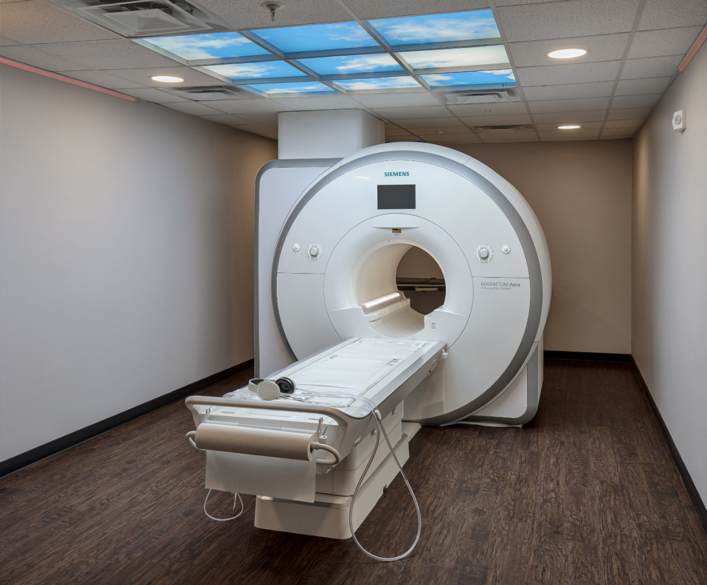
MRI
Magnetic resonance imaging (MRI) is a test that uses powerful magnets, radio waves, and a computer to make detailed pictures inside your body. Your doctor can use this test to diagnose you or to see how well you’ve responded to treatment. Unlike X-rays and CT scans, an MRI does not use radiation. Our beautiful MRI suite offers soft lighting, music of your choice, and a large opening gantry that reduces claustrophobia.
MRI exams are available by appointment: Monday 7:00 am-5:00 pm, Tuesday 7:00 am–4:00 pm, Wednesday – Friday 8:00 am-2:00pm
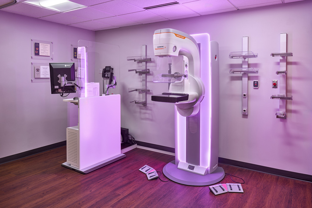
3D Mammography
Mammography is specialized medical imaging that uses a low-dose X-ray system to see inside the breasts. A mammography exam, called a mammogram, aids in the early detection and diagnosis of breast diseases in women.
Mammography is available by appointment:
Monday from 7:30 am – 5:30 pm
Tuesday from 7:30 am – 5:00 pm
Wednesday-Friday from 7:30 am – 5:30 pm
Patient Notice – Mammography (PDF)
Computed Tomography (CT)
CT (also known as CAT Scan) stands for computed tomography. Detailed images of internal organs are obtained by this type of sophisticated X-ray device. The CT scan can reveal anatomic details of internal organs, bones, tissue, and blood vessels that cannot be seen in conventional X-rays. The X-ray tube spins rapidly around the patient and the X-rays strike numerous detectors after passing through the body. These detectors are connected to sophisticated computers which generate images after image processing. The radiation dose of a CT scanner is much higher than a conventional X-ray, but the information obtained from a CT scan is often much greater and can aid your provider in making a more accurate diagnosis of your condition.
Common types of CT Scans:
Chest CT
If you are experiencing symptoms such as shortness of breath or a cough that won’t go away, your provider may order a CT of your chest. During the chest CT, an intravenous contrast may be injected to make certain features inside your chest and lungs more visible. A chest CT can help your provider diagnose lung conditions such as pneumonia, cancer, emphysema and tuberculosis.
CT Angiography
An angiogram is an X-ray image of the blood vessels used to diagnose conditions such as aneurysms (bulges in blood vessels), atherosclerosis (buildup of deposits in blood vessels), and heart abnormalities. During a CT angiogram, a dye containing iodine is injected into your bloodstream where it flows through your blood vessels and organs. As your body passes through the CT machine, the scanner collects image data from the areas highlighted by the dye and sends the information to a computer. The computer uses this information to make a 3-D picture that helps your provider make an accurate diagnosis.
Head CT
Your provider may order a head CT to help diagnose conditions such as aneurysms, brain tumors and stroke. Some head CT images are flat, while others may be 3D. In some cases, an intravenous dye contrast is injected during the scan to help highlight the inside of your head and make the CT image clearer.
Pediatric CT
Sometimes a CT scan is the only way to see inside your child’s body. We are especially careful to minimize your child’s exposure to radiation, following special procedures to determine the dose of radiation by age, weight and type of image needed. This allows us to use the least amount of radiation possible to make the highest quality images.
CT Cardiac Scoring Pricing (PDF)
CT exams are available by appointment: Monday – Friday from 7:00 am – 8:00 pm.
DEXA
Bone densitometry (DEXA) uses a very small dose of ionizing radiation to produce pictures of the inside of the body (usually the lower spine and hips) to measure bone loss. It is commonly used to diagnose osteoporosis and to assess an individual’s risk for developing fractures. DEXA is simple, quick and noninvasive. It’s also the most accurate method for diagnosing osteoporosis.
DEXA – no appointment necessary, available Monday – Friday from 7:00 am – 4:00 pm.
General Radiography
X-ray or radiography uses a very small dose of ionizing radiation to produce pictures of the body’s internal structures. X-rays are the oldest and most frequently used form of medical imaging. They are often used to help diagnose fractured bones, look for injury or infection and to locate foreign objects in soft tissue. Some x-ray exams may use an iodine-based contrast material or barium to help improve the visibility of specific organs, blood vessels, tissues, or bone.
Most x-ray exams are available 24/7.
Ultrasound
Ultrasound (also called Sonography) produces images of the inside of the body using high-frequency sound waves. Ultrasound images are captured in real-time, meaning they can also show the movement of the body’s internal organs as well as blood flowing through blood vessels. Unlike X-ray imaging, there is no ionizing radiation exposure when using ultrasound.
In an ultrasound examination, a transducer (probe) is placed directly onto the skin or sometimes into a body opening. A thin layer of gel is applied directly onto the skin allowing sound waves to be transmitted from the transducer through the gel and into the body. The transducer collects the sound that bounces back from the body and a computer uses those sound waves to create an image.
Common types of ultrasound exams:
- Abdominal: to investigate abdominal pain, nausea, vomiting, abnormal sounds, or superficial lumps.
- Pelvic: for women with pelvic pain, abnormal menstrual cycles, ovarian cysts, or other conditions associated with the female reproductive system.
- Pregnancy: used to check for fetal abnormalities, check the age and position of a fetus, and monitor fetal growth and development.
- Breast: typically used to further investigate an abnormality picked up by a provider’s physical examination or mammogram.
- Doppler: a special type of ultrasound that is used to detect the speed and direction of blood flow. This can be used in various parts of the body including neck arteries, arm arteries and veins, and leg arteries veins.
- Biopsy: ultrasound can also be used to guide procedures such as needle biopsies where small needles and/or biopsy devices are used to sample cells from an abnormal area for laboratory testing.
- Echocardiogram: A noninvasive exam using ultrasound to evaluate the heart’s function and structures. Ascension St. Vincent performs exams Monday, Wednesday and Friday from 8:00 am – 4:00 pm
Ultrasound exams are available by appointment:
Monday – Friday 8:00 am to 4:00 pm
Sneak Peek Ultrasound Packages & Pricing (PDF)
Resources
Patient Instructions on How to Access Images
Patient Instructions on How to Access Images (PDF)
Imaging Exams
3D/Tomo Screening Mammography (PDF)
3D/Tomo Screening Mammography with Breast Implants (PDF)
ABI (PDF)
Aorta Ultrasound (PDF)
Arthrogram (PDF)
Bone Density (DEXA) (PDF)
Breast Aspiration (PDF)
Breast Biopsy (PDF)
Carotid Artery Doppler (PDF)
CT Cardiac Calcium Scoring (PDF)
CT Guided Biopsy with Sedation (PDF)
CT Lung Screening (PDF)
CT with IV Contrast Prep (PDF)
CT without IV Contrast Prep (PDF)
Diagnostic Mammography (PDF)
Echocardiogram (PDF)
Entrance to Cardiac Rehab (PDF)
Esophogram/Barium swallow (PDF)
Lower Extremity Vascular Ultrasound (PDF)
Lumbar Puncture (PDF)
Modified Barium Swallow (PDF)
MRA/MRV (PDF)
MRI Exam with Contrast (PDF)
MRI Exam without Contrast (PDF)
Pregnancy Ultrasound (PDF)
Renal and Bladder Ultrasound (PDF)
Retroperitoneal Ultrasound (PDF)
Small Bowel (PDF)
Stress Treadmill (PDF)
Testicular Ultrasound (PDF)
Therapeutic Joint Injection (PDF)
Thyroid Ultrasound (PDF)
Ultrasound – Pelvic Non-OB (PDF)
Ultrasound – Pharmacology Stress Echocardiogram (PDF)
Ultrasound – Stress Echocardiogram (PDF)
Ultrasound Abdominal (PDF)
Ultrasound Abdominal Duplex (PDF)
Ultrasound Axilla (PDF)
Ultrasound Breast (PDF)
Ultrasound Chest (PDF)
Ultrasound Extremity Non-Vascular (PDF)
Ultrasound Guided Biopsy (PDF)
Ultrasound Guided Biopsy with Sedation (PDF)
Ultrasound Guided Drainage (PDF)
Ultrasound Guided Fine Needle Aspiration (PDF)
Ultrasound Guided Permanent Drain Tube – Tunnel Catheter Placement (PDF)
Ultrasound Head Neck (PDF)
Ultrasound Hernia (PDF)
Upper Extremity Vascular Ultrasound (PDF)
Upper GI – Small Bowel (PDF)
Nuclear Medicine Exams
Nuclear Medicine Bone Scan Handout (PDF)
Nuclear Medicine Cardiolite Stress Handout (PDF)
Nuclear Medicine Gastric Empty Handout (PDF)
Nuclear Medicine GI Bleed Handout (PDF)
Nuclear Medicine HIDA Scan Handout (PDF)
Nuclear Medicine I 131 Thyroid Therapy Handout (PDF)
Nuclear Medicine I-123 Thyroid Uptake Handout (PDF)
Nuclear Medicine Liver SPECT Handout (PDF)
Nuclear Medicine Liver Spleen Handout (PDF)
Nuclear Medicine Lung VQ Scan Handout (PDF)
Nuclear Medicine Meckles Handout (PDF)
Nuclear Medicine MUGA Handout (PDF)
Nuclear Medicine Parathyroid Handout (PDF)
Nuclear Medicine Renal Handout (PDF)
Hours
Although the Imaging Department remains open 24/7, examinations may need to be scheduled due to patient preps and staffing. Please contact (765) 932-7556 with any questions you may have.
Contact
Jennifer Cupp, Director of Imaging
Phone: (765) 932-7556
Fax: (765) 932-7552
Accreditations